

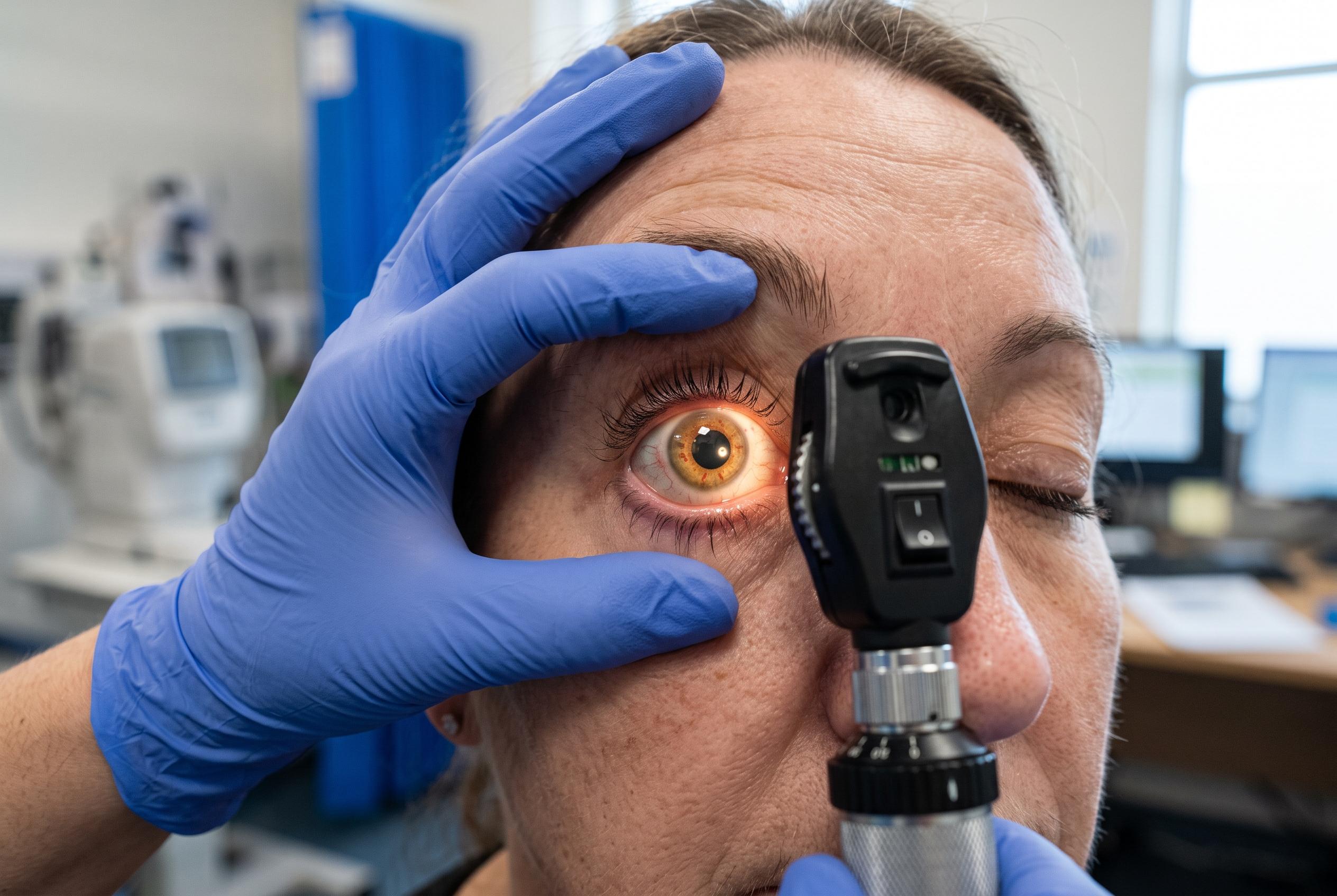

A recent review uncovers how advanced glycation end products and tissue stiffening interplay to drive the relentless progression of diabetic retinopathy, highlighting potential targets beyond traditional treatments.
Diabetic retinopathy remains one of the main causes of irreversible sight loss among working-age adults, and a review published by Dove Press argues that its progression is driven not only by biochemical injury but also by a growing mismatch between retinal metabolism and tissue mechanics. The paper says chronic hyperglycaemia promotes advanced glycation end products, or AGEs, which accumulate in the retinal microenvironment and appear to connect oxidative stress, inflammation, extracellular matrix remodelling and vascular dysfunction into one self-reinforcing process. Earlier reviews in PubMed likewise describe AGEs as central to retinal damage, including pericyte loss, microaneurysm formation, blood-retinal barrier failure and neovascularisation.
The review’s main argument is that AGEs do more than mark tissue injury. By cross-linking long-lived proteins such as collagen and elastin, they can stiffen the retinal extracellular matrix and basement membrane, making vessels less compliant and more prone to hypoxia. That stiffness then feeds back into cell signalling through mechanotransduction pathways, including integrins, Piezo1, YAP/TAZ and RhoA/ROCK. The authors say this creates a cycle in which metabolic injury is converted into mechanical stress, and mechanical stress then worsens metabolic dysfunction. A separate PubMed review from 2014 also links AGE accumulation with vascular stiffening and inflammatory signalling, supporting the broader biochemical framework on which the new paper builds.
The paper places particular emphasis on the AGE-RAGE pathway. When AGEs bind to the receptor for advanced glycation end products, downstream signalling can activate NF-kappaB, MAPK and NADPH oxidase, increasing reactive oxygen species, inflammatory cytokines and cell death. According to the review, this may help explain why retinal disease can keep advancing even after glucose control improves, because AGE-RAGE signalling may leave a lasting form of tissue “memory” in the retina. That idea is consistent with previous literature linking AGEs to chronic oxidative and inflammatory injury in diabetic retinopathy.
On the therapeutic side, the review argues that future treatment may need to go beyond anti-VEGF drugs and glucose control. It discusses several experimental approaches: blocking AGE formation with compounds such as aminoguanidine or pyridoxamine, interrupting AGE-RAGE signalling with soluble RAGE or receptor inhibitors, and trying to reverse established cross-links with AGE breakers. It also highlights targets that affect mechanical signalling, such as RhoA/ROCK, along with the possibility of reducing matrix stiffness itself by modulating lysyl oxidase. The authors caution that most of these strategies remain experimental and that human evidence is still limited.
In practical terms, the review reinforces an old message with a newer biological explanation: early screening, tight systemic risk-factor control and prompt ophthalmic care still matter most, but the retina may also be shaped by how rigid its surrounding matrix has become. The authors say that better imaging tools and stronger clinical validation will be needed before biomechanical markers or stiffness-targeting treatments can move into routine care. Until then, the work is best read as a mechanistic framework for understanding why diabetic retinopathy can be so persistent, and why it may require treatment strategies aimed at both chemistry and structure.
Disclaimer: This content is for informational purposes only and is not intended to be a substitute for professional medical judgment, advice, diagnosis, or treatment.