

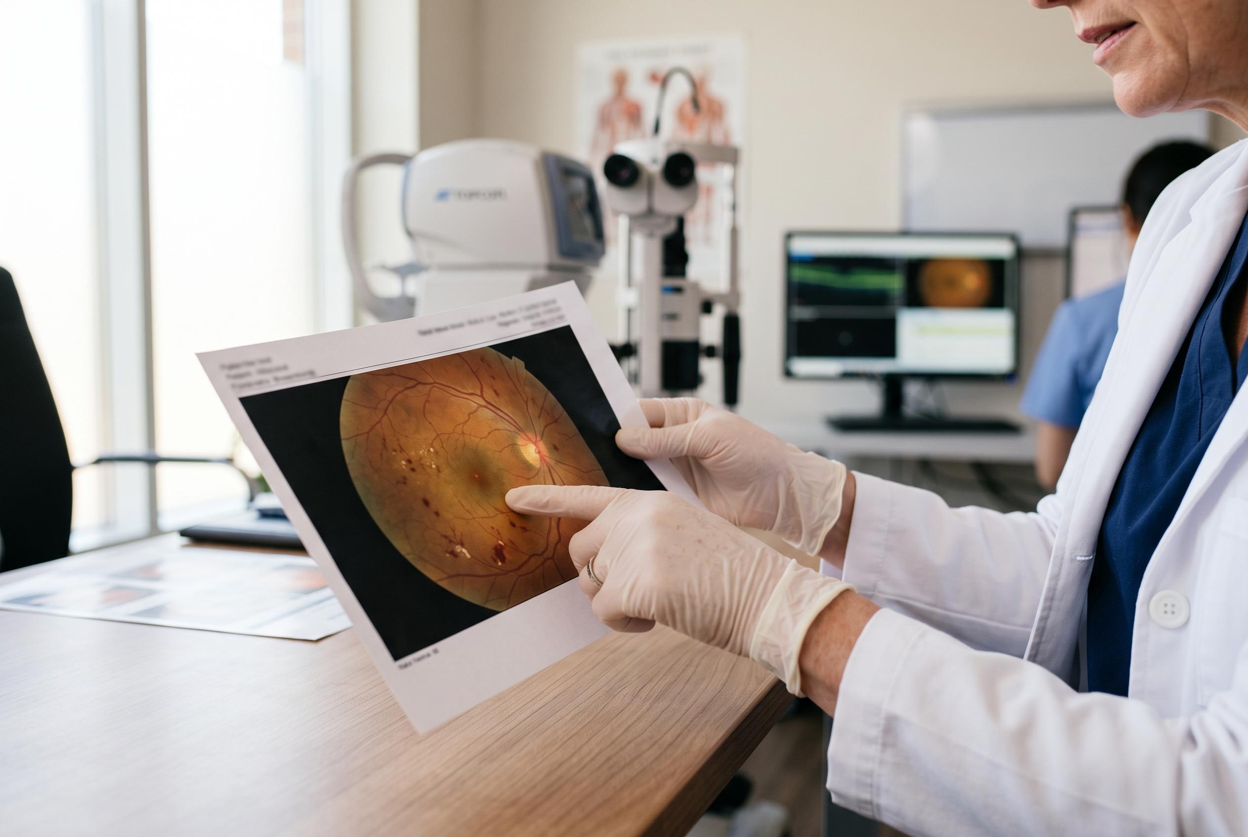

Research by Voiant and University of Turin scientists suggests pre-treatment retinal scans could help predict changes in patients’ vision and retinal thickness.
Voiant said a collaboration with researchers in Turin has published work suggesting machine-learning tools may help predict how patients with diabetic macular oedema will respond to anti-VEGF treatment. The company said the model was built to estimate both functional and structural outcomes before treatment begins, drawing on imaging data to support earlier clinical decision-making.
The paper, first authored by Lorena Ulla of Enrico Borrelli’s research group at the University of Turin and the City of Health and Science Hospital in Turin, examined whether pre-treatment scans could forecast changes in best-corrected visual acuity, or BCVA, and central subfield thickness, or CST. According to the company’s summary of the findings, the model predicted the direction of treatment response and also estimated the size of the change in vision and retinal thickness with moderate-to-strong performance.
Voiant said the study adds to its broader effort to use imaging and artificial intelligence in pre-screening and prognostic assessment. Jonathan Oakley, a senior vice-president at the company, said the firm sees prognostic imaging as a way to extend its current screening methods across more indications. Borrelli, speaking in the company’s announcement, said clinicians need as much information as possible when choosing treatment and argued that AI could help inform those decisions as well as improve the design of clinical trials.
The report lands amid growing interest in machine learning for diabetic eye disease. Recent academic work has explored unsupervised methods to sort patients with diabetic macular oedema into groups with different treatment responses, while other studies have focused on predicting persistent swelling despite therapy. Medical references describe diabetic macular oedema as a complication of diabetes in which damaged retinal blood vessels leak fluid into the macula, the part of the retina responsible for sharp central vision.
Disclaimer: This content is for informational purposes only and is not intended to be a substitute for professional medical judgment, advice, diagnosis, or treatment.