

With diabetes rising and retinal disease becoming harder to predict, Australian optometrists are being urged to combine more comprehensive imaging with closer monitoring of glucose changes, pregnancy and other high-risk factors.
Diabetic retinopathy remains one of the most important preventable causes of sight loss in Australia, and the burden is rising as diabetes becomes more common. In an article for Insight News, Royal Victorian Eye and Ear Hospital ophthalmologist Jonathan Goh said the condition is no longer something optometrists can approach as a static diagnosis, because both the risk profile and the tools available to detect it are changing quickly. The article notes that diabetes now affects well over a million Australians, with particularly high prevalence among Aboriginal and Torres Strait Islander people, and that missed screening remains a major gap in care.
That clinical gap matters because diabetic eye disease is both common and costly. The Insight News article says diabetic macular oedema alone is estimated to cost more than $2 billion a year in lost productivity and wellbeing, and that many people with diabetes still do not receive eye examinations at recommended intervals. The piece also points to Australian and New Zealand data showing that new retinopathy develops each year and that established disease can progress at widely different rates, reinforcing the need for repeated review rather than one-off assessment. That variability is one reason the article argues that optometrists remain central to early detection and referral.
The article says current Australian screening pathways recommend eye checks at diagnosis, then at least every two years when no retinopathy is present and no additional risk factors exist. Annual screening is advised when modifiers such as long diabetes duration, elevated HbA1c, uncontrolled blood pressure or lipids, pregnancy, other diabetic complications, or Aboriginal and Torres Strait Islander background are present. If retinopathy is already visible, the recommended response becomes more urgent, ranging from annual review for mild disease to same-week referral for proliferative diabetic retinopathy, with sudden visual loss requiring immediate escalation. It also notes that the relevant RANZCO guidance is under review, so local pathways should be checked regularly.
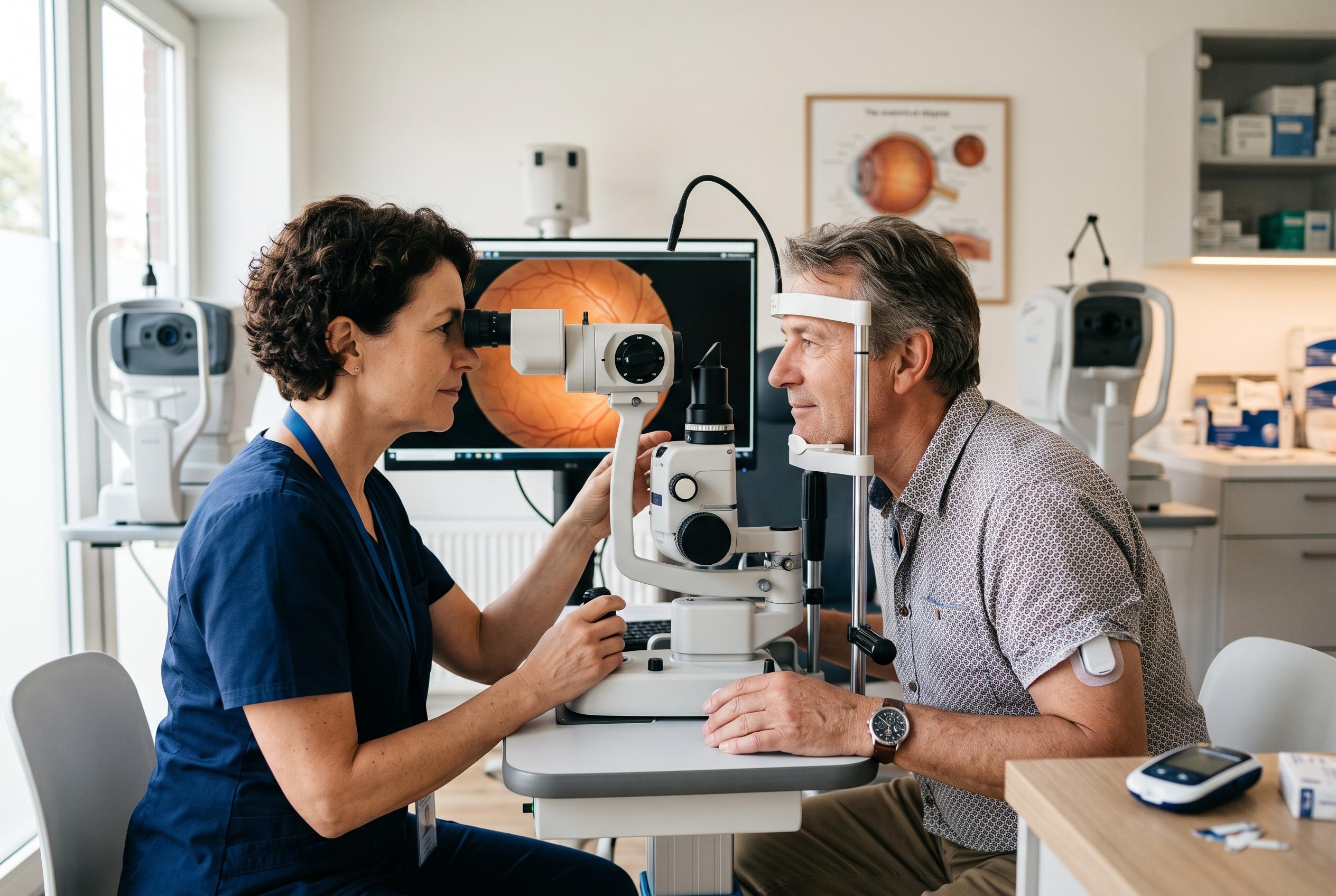
A major theme of the article is that imaging is moving beyond traditional single-field photography. Ultra-widefield imaging can capture much more of the retina than standard 45-degree images, which matters because peripheral disease may be missed if only the posterior pole is examined. A review indexed on PubMed describes ultra-widefield imaging as a valuable way to detect and monitor diabetic retinopathy and diabetic macular oedema more comprehensively, while a randomised trial found non-mydriatic ultra-widefield imaging effective for diabetic eye screening. The Insight News article says this wider view can alter severity grading and referral decisions, especially when peripheral neovascularisation is present.
Optical coherence tomography has also become indispensable for diabetic macular oedema, because it provides detailed cross-sectional views of the retina and helps clinicians judge whether treatment is needed and whether it is working. The article adds that optical coherence tomography angiography can assist in assessing macular ischaemia, while fundus fluorescein angiography remains the key test for mapping retinal ischaemia and hidden neovascular change. In combination, these tools can reveal disease that looks mild on routine examination, giving ophthalmologists a fuller picture of risk and prognosis.
The article also highlights a less widely recognised danger: retinopathy can worsen when glucose control improves rapidly. That issue has long been described with insulin, but the rise of GLP-1 receptor agonists has brought it renewed attention because these drugs can reduce HbA1c quickly. The article says a recent sharp fall in HbA1c should prompt closer surveillance, particularly in people starting insulin or a GLP-1 medicine. It also stresses that pregnancy is a separate and well-established risk period, with retinopathy sometimes accelerating during gestation and remaining active for months after delivery, which is why regular eye review through pregnancy is essential.
Continuous glucose monitoring is another development reshaping risk assessment. The Insight News article says CGM is now standard for type 1 diabetes and is expanding more broadly, while recent research indexed in PubMed and the National Library of Medicine suggests CGM-derived measures may predict retinopathy better than HbA1c alone in type 1 diabetes. Other recent studies point in the same direction, linking CGM metrics with microvascular risk and suggesting that time in range and related measures can add information that HbA1c misses. The article’s broader message is that optometrists should not rely on a single laboratory number when judging retinal risk.
Treatment has also advanced. The article says pan-retinal photocoagulation remains the backbone of care for proliferative disease, but anti-VEGF injections are now used more often, including as a bridge when laser cannot be completed immediately. For diabetic macular oedema, anti-VEGF agents remain first-line, while steroid implants and focal or grid laser still have selected roles. The practical message, the article concludes, is unchanged even as the technology evolves: recognise rising risk early, monitor closely, and refer before sight is lost.
Disclaimer: This content is for informational purposes only and is not intended to be a substitute for professional medical judgment, advice, diagnosis, or treatment.