

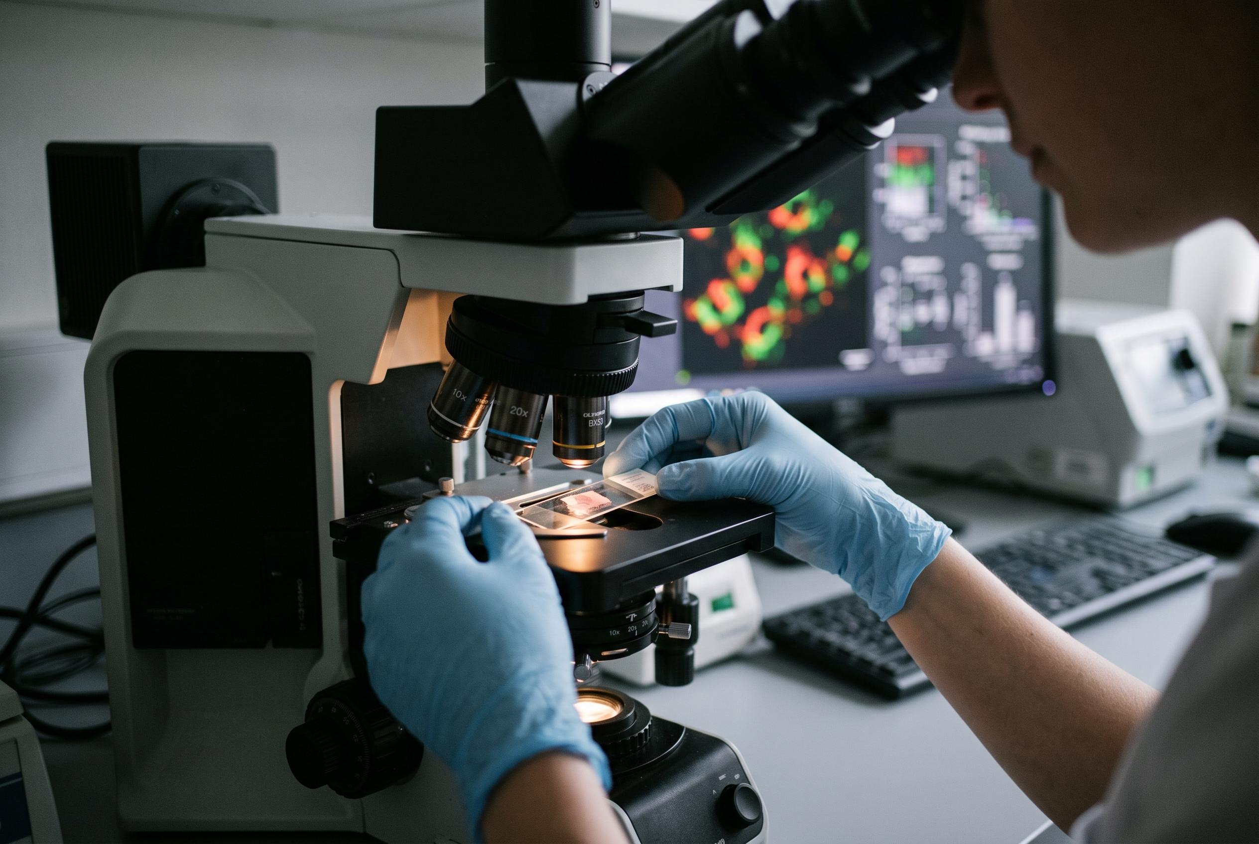

A recent human tissue study highlights reduced expression of mitophagy-related genes, especially PINK1 and ULK1, in diabetic kidney disease, suggesting new avenues for diagnosis and treatment.
A new study in Scientific Reports adds to growing evidence that disrupted mitophagy,the process cells use to clear damaged mitochondria,may be part of the biology behind diabetic nephropathy, a serious kidney complication of diabetes. In renal cortical biopsy samples from patients with advanced disease, the researchers found lower expression of several genes involved in the AMPK-ULK1-PINK1-Parkin pathway, while comparing the findings with control tissue taken from non-diseased regions of nephrectomy specimens. (pubmed.ncbi.nlm.nih.gov)
The team analysed 66 samples in total, split evenly between people with diabetic nephropathy and controls, and measured mitophagy-related mRNA in kidney tissue using quantitative PCR. According to the study summary, patients with diabetic nephropathy also showed worse metabolic profiles, including higher glucose, HbA1c, insulin resistance and lipid values, alongside lower indices associated with insulin sensitivity. Several genes, including PRKAA1, ULK1, PINK1, MAP1LC3B and BECN1, were significantly reduced, while PRKN, SQSTM1 and MTOR did not differ meaningfully between the groups. (pubmed.ncbi.nlm.nih.gov)
The most striking exploratory signals came from PINK1 and ULK1, which showed the strongest discrimination between cases and controls in receiver-operating analyses, with area-under-the-curve values of 0.762 and 0.748 respectively. The study also reported correlations between gene expression and measures of kidney function such as estimated glomerular filtration rate and creatinine, although the transcripts did not track neatly with histological severity. That fits with earlier work pointing to ULK1 as a potential biomarker in diabetic nephropathy, including a 2022 analysis that found the gene reduced in glomerular tissue from patients with the condition and linked it to kidney function. (pubmed.ncbi.nlm.nih.gov)
Taken together, the findings strengthen a broader body of research suggesting that impaired mitophagy can contribute to mitochondrial dysfunction, inflammation and fibrosis in diabetic kidney disease. Earlier experimental studies have already implicated the PINK1-Parkin axis, while other work has shown that manipulating related pathways, including mTOR and TXNIP, can restore autophagic flux in diabetic kidneys in preclinical models. The new study does not prove causation, but it adds human tissue evidence to a field that has until now relied heavily on animal and bioinformatic data. The authors said larger and more clinically diverse cohorts will be needed before the results can be translated into practice. (pubmed.ncbi.nlm.nih.gov)
Disclaimer: This content is for informational purposes only and is not intended to be a substitute for professional medical judgment, advice, diagnosis, or treatment.