

Ultra-widefield imaging, OCT and continuous glucose monitoring are reshaping how Australian optometrists detect and manage diabetic eye disease before vision is lost.
Diabetic retinopathy is becoming harder to ignore as diabetes continues to affect more people and the burden on eye services grows. In Australia, the condition remains a major cause of preventable vision loss, and Royal Victorian Eye and Ear Hospital ophthalmologist Jonathan Goh argues that optometrists are increasingly central to early detection, surveillance and referral. He points to advances in imaging, changing treatment options and a better appreciation of clinical risk factors as reasons the approach to diabetic eye disease is evolving.
The scale of the problem is substantial. The article notes that diabetes affects well over a million Australians, with prevalence rising alongside ageing and higher rates of obesity. It cites the National Eye Health Survey, which found self-reported diabetes in about 14% of non-Indigenous older adults and 36% of Indigenous older adults. Among Australians over 50, diabetic retinopathy is associated with visual impairment in roughly one in 25 non-Indigenous people and up to one in six Indigenous people. A recent meta-analysis of Australian and New Zealand cohorts also found that new retinopathy developed at an annual rate of 4.22%, while progression among those already affected ranged from 3.08% to 18.22% a year.
That variability matters because diabetic retinopathy is not a stable diagnosis. The article says the economic burden is also considerable, with diabetic macular oedema estimated to cost Australians more than $2 billion a year in lost productivity and wellbeing, affecting around 72,000 people. It adds that about half of people with diabetes are not getting eye examinations at recommended intervals, and modelling suggests DMO prevalence could rise sharply if screening gaps persist. That makes routine optometric review one of the most effective interventions available.
The pathophysiology remains familiar, but the practical lesson is that damage begins long before vision changes. Chronic hyperglycaemia injures retinal microvessels, weakening the blood-retinal barrier and leading to haemorrhages, lipid leakage and capillary loss. As oxygen supply drops, the retina increases production of growth factors such as vascular endothelial growth factor, or VEGF, which drives leakage, swelling and the growth of fragile new vessels. In other words, the disease progresses from early microvascular injury to macular oedema and, in advanced cases, proliferative retinopathy.
For Australian optometrists, the RANZCO screening and referral pathway remains the main framework, although the article notes that it is under review. Patients with diabetes should be screened at diagnosis and then at least every two years if there are no retinopathy signs or risk modifiers. Annual review is recommended for people with longer diabetes duration, poor glycaemic control, hypertension, dyslipidaemia, other diabetic complications, pregnancy or Aboriginal and Torres Strait Islander background. Once retinopathy appears, the referral timeline shortens quickly: mild non-proliferative disease can be reviewed in a year, moderate disease should be referred within 12 weeks, severe disease within four weeks, proliferative disease within one week, and sudden visual loss needs same-day ophthalmic assessment. The article also notes that Victorian public hospital thresholds are stricter, with referrals generally reserved for severe non-proliferative disease, proliferative disease and centre-involving macular oedema with reduced vision.
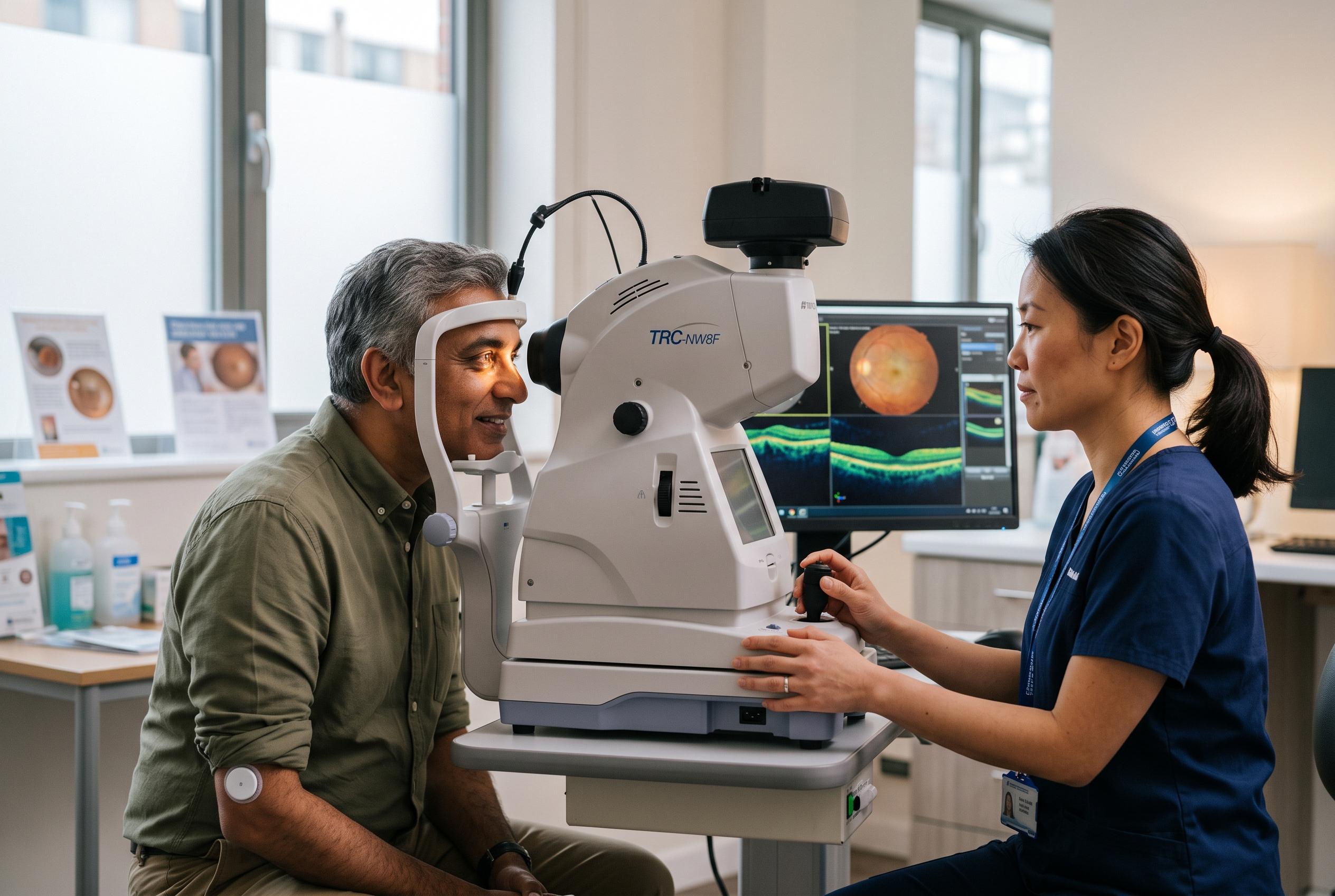
Imaging is where practice has changed most. The article argues that a single 45-degree photograph is no longer enough for screening because much of the retina is left unseen. Ultra-widefield imaging can capture as much as 200 degrees of retina and may reveal peripheral disease that would otherwise be missed, including neovascularisation. A review on ultra-widefield imaging in diabetic eye disease likewise concludes that it improves visualisation of peripheral lesions and can influence staging and treatment planning. Optical coherence tomography has become essential for assessing diabetic macular oedema because it provides cross-sectional detail that helps quantify swelling and monitor response to treatment. Fundus fluorescein angiography remains important for judging retinal ischaemia, which is often the key driver of sight-threatening disease.
The article also highlights a less familiar risk: retinopathy can worsen when glucose control improves rapidly. That phenomenon was first linked to insulin therapy, but it has become more visible again with the wider use of GLP-1 receptor agonists such as semaglutide, marketed as Ozempic and Wegovy. A sharp fall in HbA1c can be followed by temporary worsening of retinopathy, including progression to proliferative disease, so a recent dramatic improvement in glycaemic control should prompt closer observation. Pregnancy is another well-established trigger for faster progression, with risk influenced by baseline disease, longer diabetes duration, rapid HbA1c reduction and conditions such as hypertension or pre-eclampsia. The article says pregnant women with pre-existing diabetes need retinal review in the first trimester, again at 28 weeks if no retinopathy is present, and earlier repeat assessment if eye disease is already present.
Continuous glucose monitoring is emerging as an additional tool in risk assessment. The article says CGM is now standard in type 1 diabetes and increasingly used more broadly, with Australian uptake rising sharply among hospital inpatients with type 1 diabetes in Melbourne. Importantly, it says CGM-derived average glucose may predict proliferative retinopathy better than HbA1c alone. That suggests optometrists should look beyond the traditional laboratory marker and consider CGM data, especially in type 1 diabetes, as part of a fuller picture of retinal risk.
Treatment options have also expanded. For proliferative diabetic retinopathy, pan-retinal photocoagulation remains the cornerstone because it reduces the ischaemic drive by treating peripheral hypoxic retina. The article says ranibizumab, now subsidised in Australia for proliferative disease, may be used to stabilise eyes or bridge the period before laser can be completed, but it requires careful follow-up because missed injections can lead to rebound disease. For diabetic macular oedema, anti-VEGF injections remain first-line, while corticosteroid implants are generally reserved for cases that do not respond adequately, given the risk of cataract and raised intraocular pressure. Focal or grid laser still has a role in selected non-centre-involving cases, and vitrectomy is reserved for non-clearing vitreous haemorrhage or traction threatening the macula.
The article’s broader message is straightforward: diabetic eye disease is more detectable, more measurable and more treatable than it once was, but only if clinicians act early. Better imaging, more detailed glucose data and new therapies are improving care, yet the fundamentals still depend on timely screening, careful follow-up and prompt referral before sight is lost.
Disclaimer: This content is for informational purposes only and is not intended to be a substitute for professional medical judgment, advice, diagnosis, or treatment.